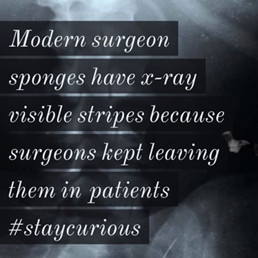
Modern surgeon sponges have x-ray visible stripes because surgeons kept leaving them in patients

Gossypiboma is the official name for a retained sponge/towel after surgery. This word comes from the Latin word for cotton, gossypium, combined with the Swahili word for place of concealment, boma.[7] It is also commonly referred to as textilioma. This word combines textile, meaning cloth, and the suffix –oma, which means growth or tumor.[7]A case of gossypiboma can be subtle and may not be discovered until months or even years after the surgery has been performed. In rare cases, a situation can be so severe that it is noticed immediately. Some of the ways gossypiboma can present itself are as a mass in the body or as a bowel tumor.[8] Immediately after surgery, a case of gossypiboma can commonly be mistaken for an abscess, especially when it is near a passage between organs (a ‘fistula’). In those cases where a sponge isn’t discovered until much later, it may be impossible to tell the difference between gossypiboma and an ‘intra-abdominal abscess’.[7] This is because both cause air bubbles and “calcification of the cavity wall.”[7] Gossypiboma is difficult to diagnose due to vague, inconsistent symptoms and images from x-rays that provide no solid evidence and unclear results. Because it is difficult to diagnose, emphasis has been put on the prevention of the mistake. The following techniques have been put into practice to prevent gossypiboma. Radiopaque marking: Before operation, sponges can be soaked through with ‘radio-opaque marker’. This allows a sponge to be easily seen on plain radiographs. When the markers are noticed, it can be assumed that it is revealing a retained sponge. A.P. Zbar, Surgical Directorate at Oldchurch and Harold Wood Hospitals stated “the diagnosis is easily made by plain abdominal radiography, when a radio-opaque marker is seen”.[8] This method is flawed in that it doesn’t work if the sponges have broken into smaller pieces over time.
Ultrasonography- Gossypiboma can be recognized with ultrasonography by “the presence of brightly echogenic wavy structures in a cystic mass showing posterior acoustic shadowing that changes in parallel with the direction of the ultrasound beam” according to Zbar [8] and associates.
Computerized Tomography (CT)- A surgical sponge on a CT will show air bubbles on soft tissue masses. The flaw with this technique is that gossypibomas are easily confused with abscesses.
@Curionic
#staycurious